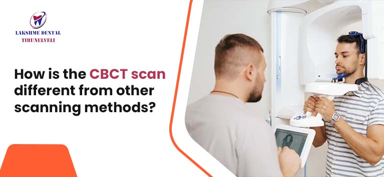
How is the CBCT scan different from other scanning methods?
Establishing a precise diagnosis in odonto-stomatology requires a careful clinical examination, often supplemented by additional radiological examinations. Cone beam or CBCT (Cone Beam Computed Tomography), also called cone beam digital volume tomography, is a booming 3D sectional imaging technique. It allows the exploration of calcified tissues, namely bone and teeth, with unparalleled precision and minimal radiation exposure, making it a go-to choice in various fields. In this article, you will delve into the unique attributes of CBCT and explore how it differs from conventional scanning techniques.
What Is CBCT?
CBCT, or Cone Beam Computed Tomography, is a radiographic examination tool that provides 3D images of the patient’s mouth.
It allows us to see practically perfectly any morphological variety in the teeth and surrounding tissue, such as bone and nerve structures.
Likewise, it is of great help in discovering injuries of all kinds that are related to the bone, whether cysts, granulomas, or fractures.
Furthermore, it is a very practical tool to detect possible failure in Endodontics due to its ability to see how the canals have been treated and the length that the filling can reach. In this way, a better assessment of the case can be made and thus diagnose a prognosis with greater accuracy.
All of this can be carried out thanks to the three dimensions of the images obtained, allowing us to reach areas that are normally hidden in a conventional X-ray.
What Uses Does CBCT Have?
CBCT equipment can be divided according to the volume of the image or field of view (or, as it is called, Field of View in English). This tool can be of large FOV, that is, 6 to 12 inches or 15-30.5 cm. or tools with limited FOV, or in other words, from 1.6 to 3.1 inches or 4 to 8 cm.
It could be said that the larger the FOV, the greater the represented extension of the anatomical part, and, therefore, the greater the patient’s radiation exposure and the lower its image resolution.
Normally, the CBCT scanner is used to solve problems caused by Orthodontics; however, it is also very beneficial in more difficult scenarios such as:
- The exact location of a dental implant
- Study and planning before performing surgery
- Know what the origin of pain is
- Complete cephalometric examination
- Define oral bone structures and tooth positioning
- Diagnosis of Temporomandibular Joint Disorders (TMJ, facilitating tailored treatments)
- Carry out a good evaluation study of the jaws, paranasal sinuses, nerve channels, and nasal cavity
- Early detection and management of jaw tumours
- Perform reconstructive surgery
What Benefits Does CBCT Have?
The CBCT scanner brings us multiple benefits compared to other techniques. Some of the advantages that we can point out from a CBCT scan:
- It has a very high image scanner quality
- This is a quick practice, completed in approximately 2 minutes
- The scanner takes images from many angles, which can be manipulated digitally, allowing for a more complete evaluation
- The radiation that the patient must undergo is very low. The dose of a conventional CT scan is the same as that received in 75 CBCT
- After the technique, the radiation does not remain in the patient’s body
- Soft tissue and bone tissue can be observed at the same time
- It allows radiolucent endodontic lesions to be detected before they can be detected on conventional radiographs
- It is a painless and non-invasive technique, and it does not cause any side effects
- The patient can be scanned sitting instead of lying down, thus achieving a study with the patient’s natural head position
- It is an open scanner, so it provides greater comfort and avoids possible claustrophobic situations
What Are the Differences between CT And 3D CT or CBCT?
The big difference to highlight between the two is the degree of radiation to which the patient is subjected. The level of radiation emitted with CBCT is much lower than with CT.
Additionally, CBCT scans are notably faster, with results available in just two minutes. Therefore, it has been a great advance for dentists since it allows them to make a practically immediate diagnosis.
The information obtained is collected in the DICOM format that allows advanced processing with specific software for both visualization and therapeutic planning. The image obtained is isotropic; that is, the voxel, the minimum unit of information – equivalent to the pixel in photography – has the same dimensions in the three axes of space, thus allowing the image obtained not to be distorted.
Finally, in CBCT, the patient is scanned standing instead of lying down, as is done in CT. In this way, we manage to carry out an examination in the natural position of the head. Another advantage of CBCT, as already mentioned, is that it is an open scanner, so possible problems of claustrophobia are avoided.
Conclusion
In summary, it can be determined that three-dimensional technology is a field that has been booming in recent years, and it is essential to investigate possible applications of new devices to make more accurate diagnoses and treatments and thus obtain better results. The CBCT scan stands apart from other Scanning Methods due to its ability to provide high-precision 3D imaging with reduced radiation exposure. CBCT has transformed diagnostics and imaging by providing real-time imaging, increased soft tissue visibility, and decreased artifacts, ushering in a new era of precision and efficiency in healthcare and beyond. As technology continues to advance, the CBCT scan will undoubtedly play an increasingly pivotal role in various domains, reaffirming its status as a game-changer in the world of imaging and diagnostics. If you need to undergo any type of scan, contact a professional dental clinic to undergo a high-precision study.


